
The Neuron Poster
Source-backed
Every label has a source. Short citations are printed on the poster; the full list is below.
Drawn by code, not AI
The anatomy is generated as vector geometry from code. No AI imagery.
Readable labels
Structure names about 17 pt; no label text under 12 pt at 18 × 24 in.
Details
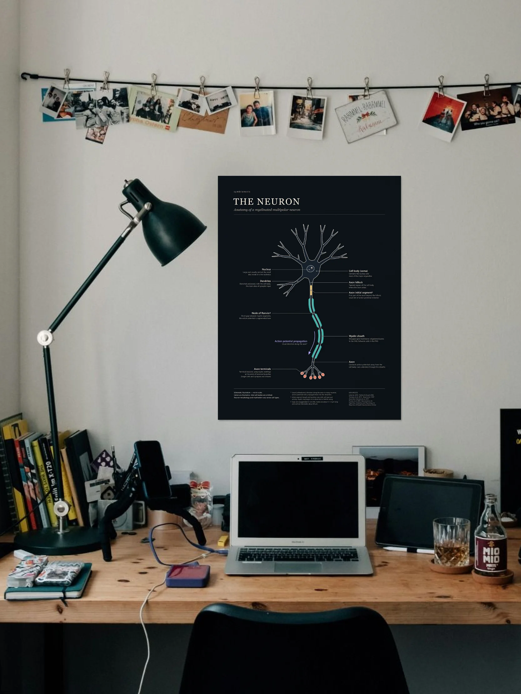
18 × 24 in (45.7 × 61 cm), portrait. Fits a standard 18 × 24 in frame. Frame not included.
Materials
Enhanced matte paper, 189 g/m² (0.26 mm). Water-based inkjet print, no glare.
Shipping & returns
Printed to order and shipped rolled; arrives in about 5–11 days. Shipping is calculated at checkout. See Returns & FAQ for damaged prints.
Up close
What's on the poster
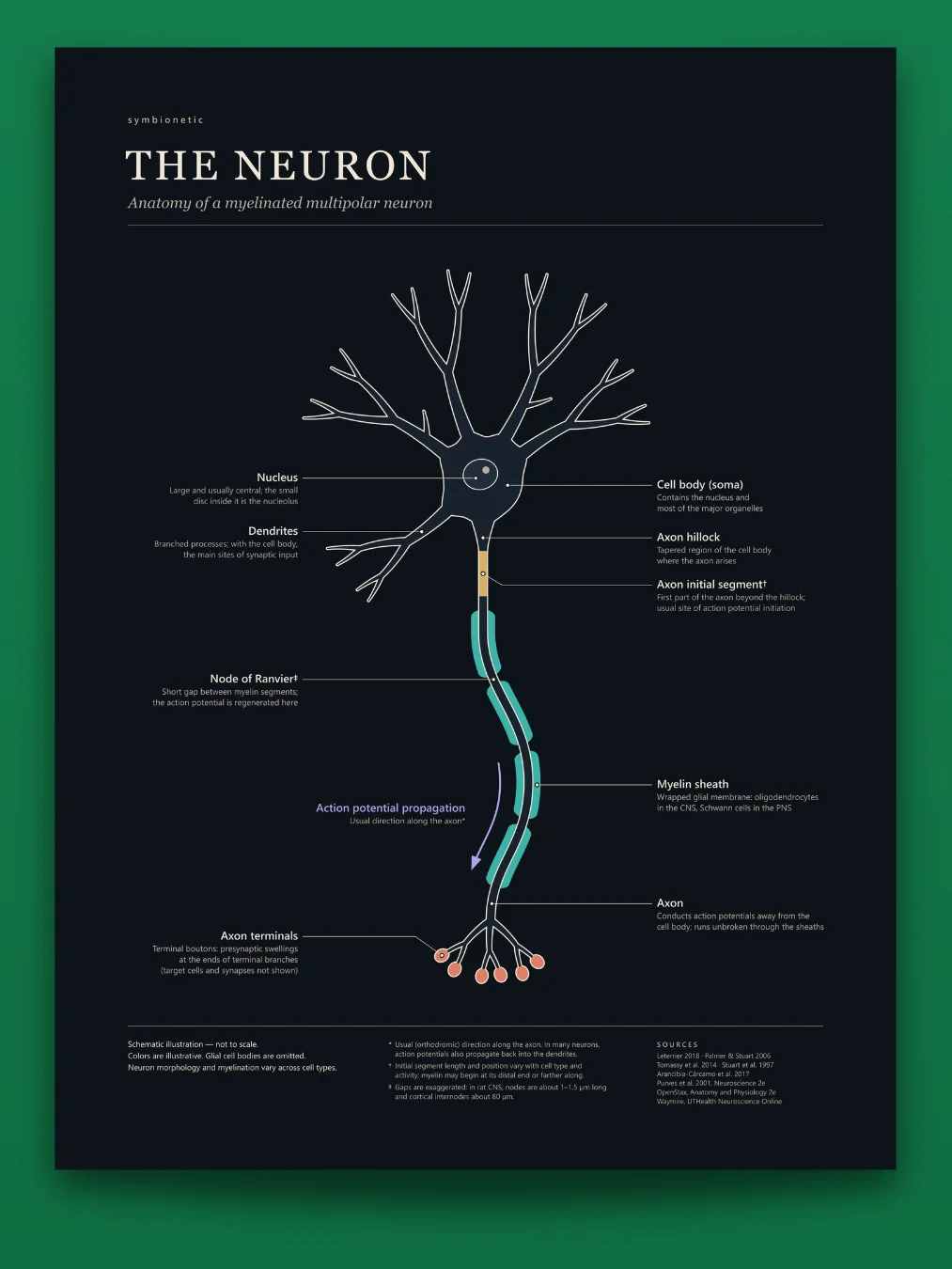
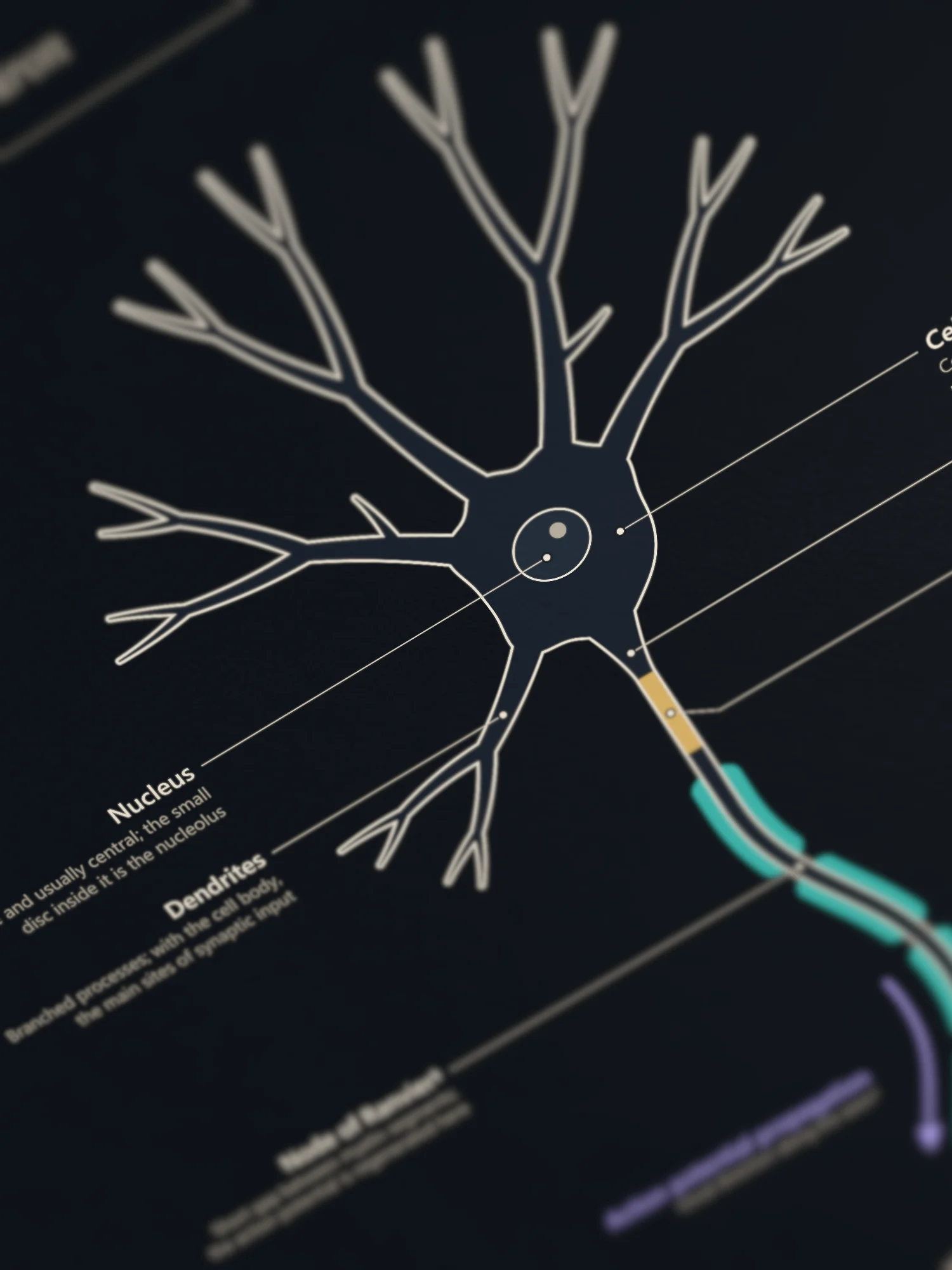
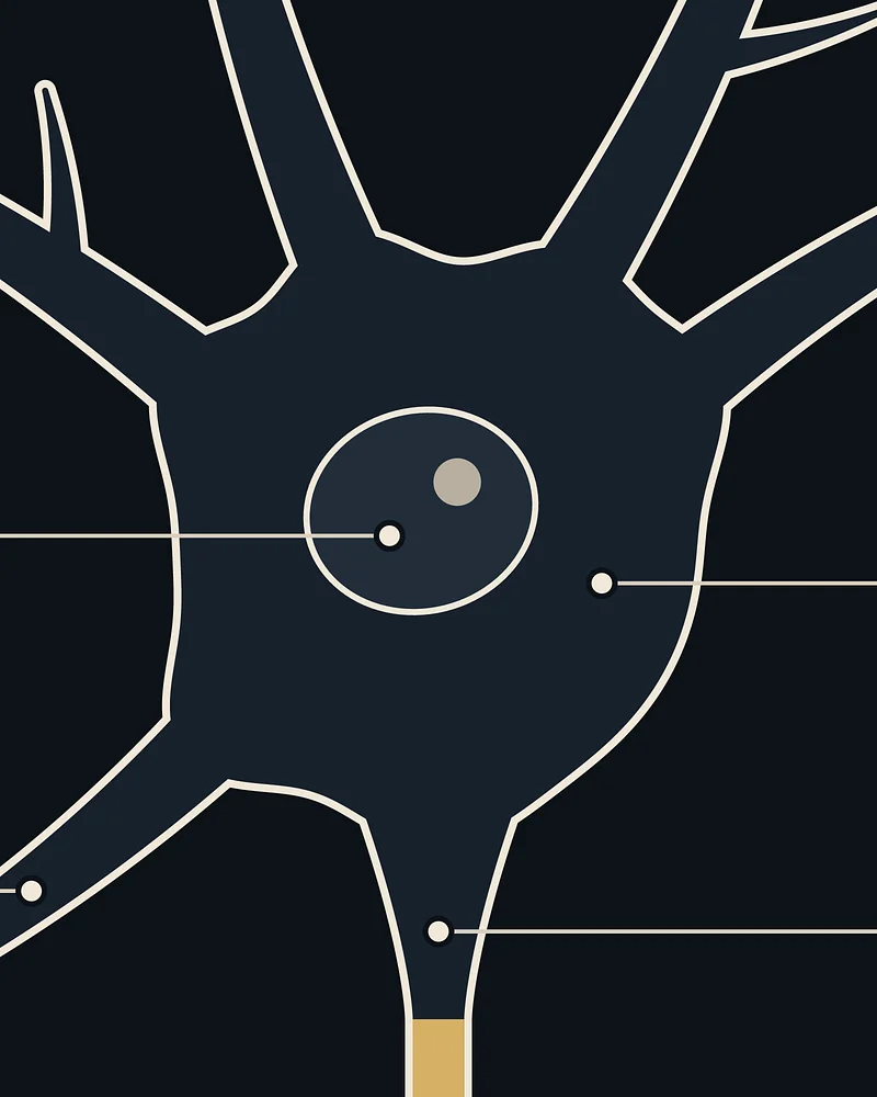
- Dendrites: branched processes; with the cell body, the main sites of synaptic input
- Nucleus: large and usually central; the small disc inside it is the nucleolus
- Cell body (soma): contains the nucleus and most of the major organelles
- Axon hillock: tapered region of the cell body where the axon arises
- Axon initial segment: first part of the axon beyond the hillock; usual site of action potential initiation
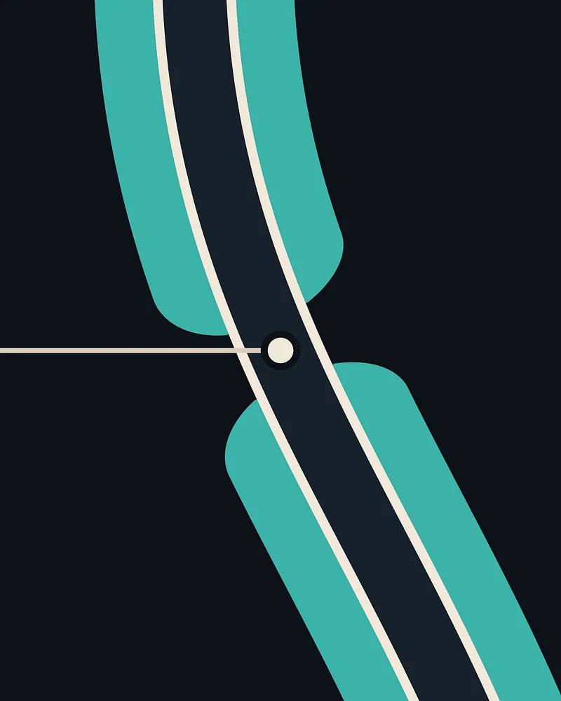
- Axon: conducts action potentials away from the cell body; runs unbroken through the sheaths
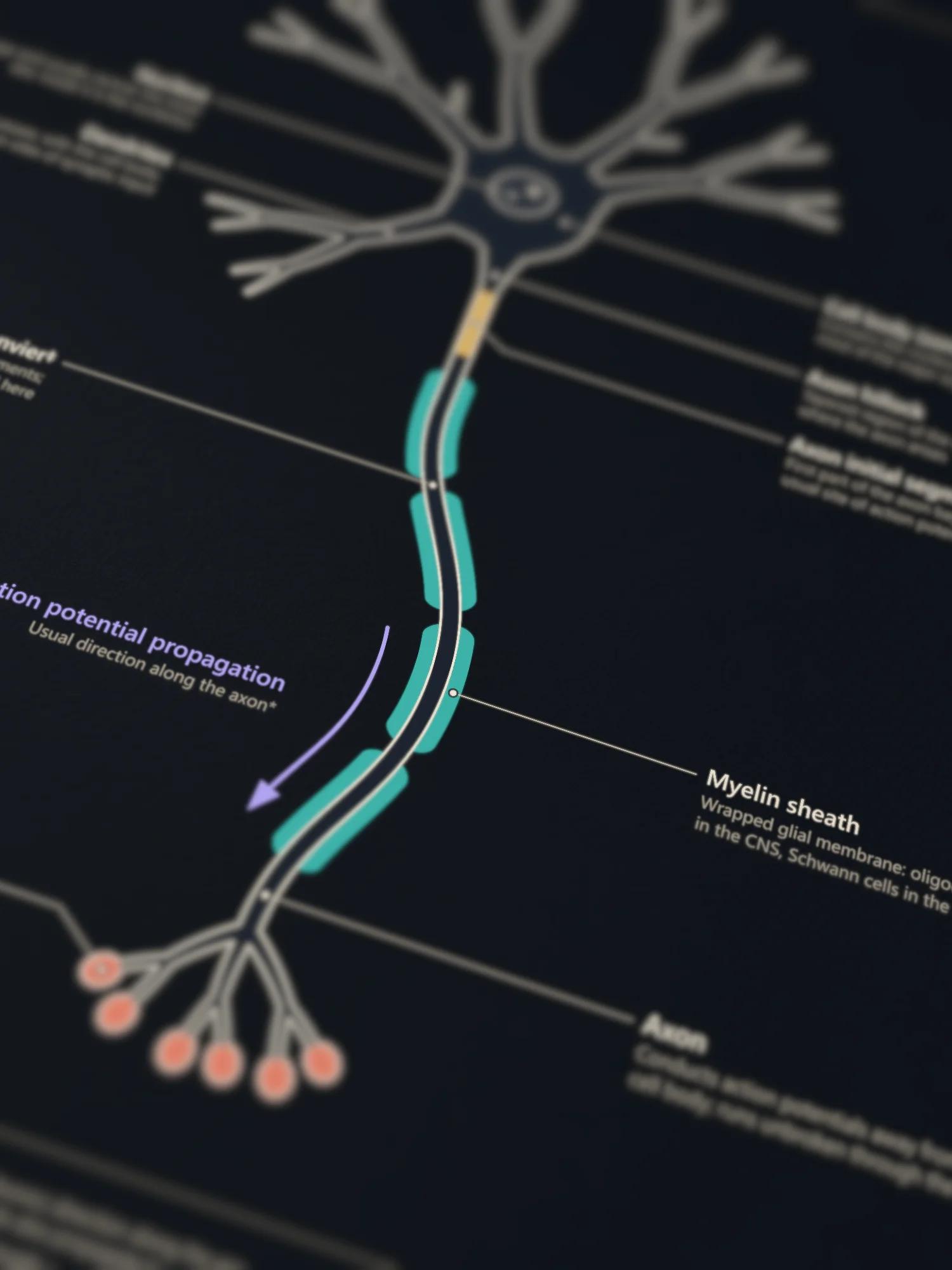
- Myelin sheath: wrapped glial membrane: oligodendrocytes in the CNS, Schwann cells in the PNS
- Node of Ranvier: short gap between myelin segments; the action potential is regenerated here
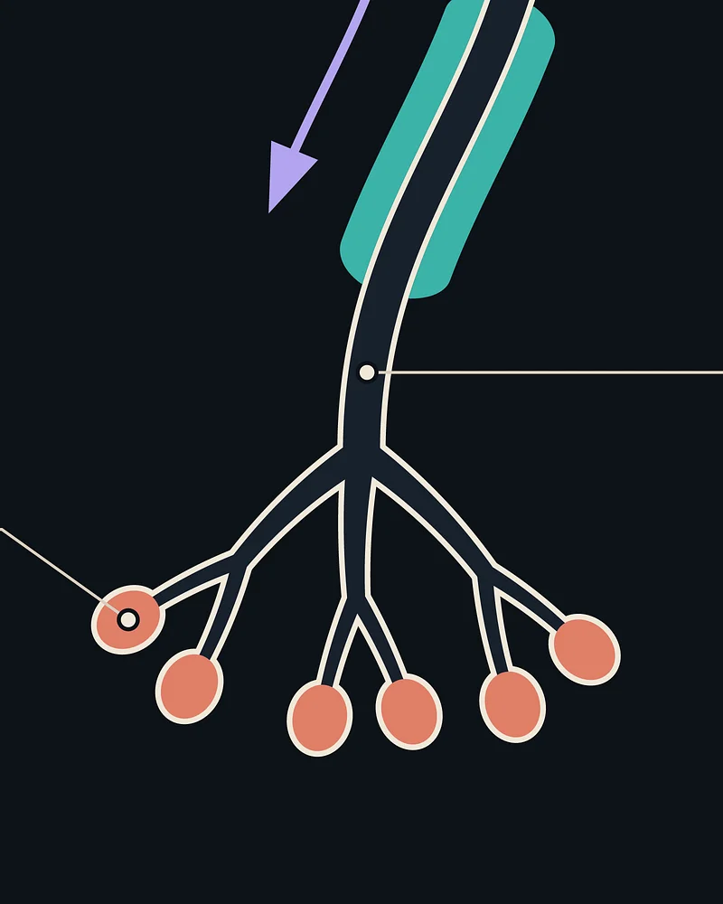
- Axon terminals: terminal boutons, the presynaptic swellings at the ends of terminal branches (target cells and synapses not shown)
- Action potential propagation (arrow): usual direction along the axon
* Usual (orthodromic) direction along the axon. In many neurons, action potentials also propagate back into the dendrites.
† Initial segment length and position vary with cell type and activity; myelin may begin at its distal end or farther along.
‡ Gaps are exaggerated: in rat CNS, nodes are about 1–1.5 µm long and cortical internodes about 80 µm.
Sources & accuracy
A schematic, not to scale. Colours are illustrative, glial cell bodies are omitted, and neuron shape and myelination vary between cell types.
What we simplified
- Dendrites: spines are not drawn; the number of dendrites and their branching are representative.
- Cell body and nucleus: organelles (Nissl bodies, Golgi apparatus, mitochondria) and the nuclear envelope with its pores are not drawn; sizes are not to scale.
- Axon hillock and initial segment: the initial segment is labelled as the usual site where the action potential starts. Textbooks differ here: Purves et al. (2001) name the hillock, and OpenStax counts the hillock and initial segment together as the trigger zone.
- Axon: one axon without collaterals; real axons range from micrometres to about a metre in length.
- Myelin: sheaths are drawn evenly spaced and of equal length, though real sheaths vary; along some cortical axons myelin is patchy. The glial cells that form myelin are not drawn.
- Nodes of Ranvier: the gaps are enlarged so they are visible (‡).
- Axon terminals: the number and shape of boutons are representative; vesicles, the synaptic cleft and target cells are not drawn.
- Arrow: shows the usual direction only (*). Saltatory, node-to-node conduction is not drawn.
References
Sources 1–8 are printed in short form on the poster.
Primary literature
- Leterrier C. (2018). The axon initial segment: an updated viewpoint. Journal of Neuroscience 38(9):2135–2145. Link
- Palmer L. M., Stuart G. J. (2006). Site of action potential initiation in layer 5 pyramidal neurons. Journal of Neuroscience 26(6):1854–1863. Link
- Tomassy G. S., et al. (2014). Distinct profiles of myelin distribution along single axons of pyramidal neurons in the neocortex. Science 344(6181):319–324. Link
- Stuart G., Spruston N., Sakmann B., Häusser M. (1997). Action potential initiation and backpropagation in neurons of the mammalian CNS. Trends in Neurosciences 20(3):125–131. Link
- Arancibia-Cárcamo I. L., et al. (2017). Node of Ranvier length as a potential regulator of myelinated axon conduction speed. eLife 6:e23329. Link
Textbooks
- Waymire J. C. Organization of Cell Types (Section 1, Chapter 8). Neuroscience Online, UTHealth. Link
- OpenStax. Anatomy and Physiology 2e: 12.2 Nervous Tissue · 12.4 The Action Potential
- Purves D., et al. (2001). Neuroscience, 2nd ed. Sinauer Associates: Nerve Cells · Increased Conduction Velocity as a Result of Myelination
Also consulted
- Purves D., et al. (2001). Neuroscience, 2nd ed. Neuroglial Cells
- Waters J., Larkum M., Sakmann B., Helmchen F. (2003). Supralinear Ca²⁺ influx into dendritic tufts of layer 2/3 neocortical pyramidal neurons in vitro and in vivo. Journal of Neuroscience 23(24):8558–8567. Link
How it's made
Every structure on this poster is drawn as vector geometry by code, checked against the sources above, and verified automatically for type sizes, text overlaps, leader lines and PDF fonts. No AI-generated images.